

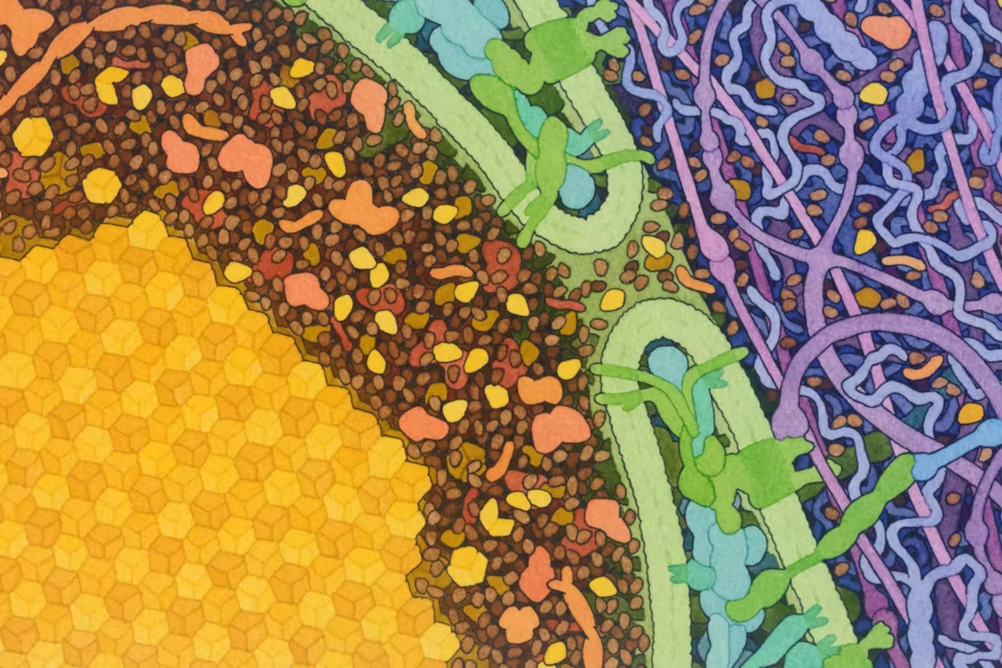
Every human being has more than 20,000 proteins. For example, hemoglobin, which transports oxygen from the lungs to cells throughout the body, or insulin, which signals to the body that sugar is present in the blood.
Each protein is made up of a sequence of amino acids, the order of which determines its folding and spatial structure—much as a word folds in space depending on the sequence of letters that make it up. This sequence and the protein’s folding (or structure) determine its biological function: the study of these is the field of “structural biology.” It relies on various complementary experimental methods, which have led to considerable advances in our understanding of the living world in recent decades and, in particular, enable the development of new drugs.
Since the 1970s, researchers have been trying to determine protein structures based solely on knowledge of the amino acid sequence (a process known as “ab initio”). It was only very recently, in 2020, that this became possible on a near-systematic basis, withthe rise of artificial intelligence and, in particular, AlphaFold, an AI system developed by a Google-owned company.
Given these advances in artificial intelligence, what is the role of structural biologists now?
To understand this, it is important to recognize that one of the challenges facing biology in the future is “integrative biology, ” which aims to understand biological processes at the molecular level within their cellular contexts. Given the complexity of biological processes, a multidisciplinary approach is essential. It relies on experimental techniques, which remain indispensable for studying protein structure, dynamics, and interactions. Furthermore, each of these experimental techniques can benefit in its own way from AlphaFold’s theoretical predictions.
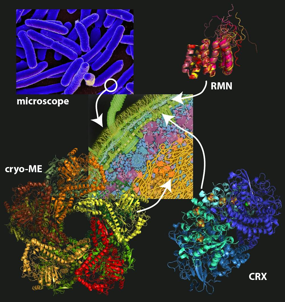
As of now, crystallography is the most widely used technique in structural biology. It has made it possible to catalog more than 170,000 protein structures in the “Protein Data Bank,” representing more than 10,000 different folds.
[Nearly 80,000 readers rely on The Conversation’s newsletter to better understand the world’s major issues. Subscribe today]
To use X-ray crystallography, proteins must be “crystallized.” It is often said that this technique is limited by the quality of the protein crystals, which is lower for large proteins. But this notion does not always reflect reality: for example, the structure of the ribosome—the enormous molecular machine that assembles proteins—was determined at a resolution of 2.8 angstroms. Venkatraman Ramakrishnan, Thomas Steitz, and Ada Yonath were awarded the Nobel Prize in Chemistry in 2009 for this work.
With the recent development of the free-electron X-ray laser (XFEL), it has become possible to simultaneously study thousands of protein microcrystals at room temperature and on the femtosecond timescale (10⁻¹⁵ seconds, or one millionth of a billionth of a second—the timescale on which chemical reactions and protein folding occur). This technique allows scientists to image proteins before they are destroyed. It is revolutionizing “kinetic crystallography, ” which makes it possible to see proteins “in action,” as well as drug discovery.
So far, AlphaFold’s contribution to the study of protein structure through crystallography has focused on generating protein models that are accurate enough to apply the technique known as “molecular replacement” to structure determination.
Another experimental method for studying protein structure is “nuclear magnetic resonance spectroscopy.” While its counterpart in medical imaging, MRI, examines the spatial distribution of a single signal—characteristic of the chemical elements in the biological tissues being observed—in nuclear magnetic resonance spectroscopy, it is a set of signals from the atoms that make up the protein that is recorded (known as the “spectrum”).
Generally, structure determination by magnetic resonance is limited to proteins of modest size. Molecular models are calculated based on structural parameters (such as interatomic distances) derived from the analysis of experimental spectra. This can be thought of as similar to the early days of cartography, when distances between reference points were used to draw 2D maps. To make it easier to interpret spectra that contain a great deal of information, models obtained through prediction (rather than experimentally) can be used, as with AlphaFold.
In addition to structural determination, nuclear magnetic resonance spectroscopy offers two major advantages. First, the analysis is generally performed on a sample in aqueous solution, making it possible to observe particularly flexible regions of proteins that are often invisible using other techniques. It is even possible to quantify their motion in terms of amplitude and frequency, which is extremely useful because the internal dynamics of proteins are just as crucial to their function as their structure.
Furthermore, nuclear magnetic resonance spectroscopy makes it easy to detect interactions between proteins and small molecules (ligands, inhibitors) or other proteins. This allows for the identification of interaction sites, information that is essential, among other things, for the rational design of active molecules such as drugs.
These properties make nuclear magnetic resonance spectroscopy an extraordinary tool for the functional characterization of proteins, in conjunction with other experimental techniques and AI.
Cryo-electron microscopy involves rapidly freezing (to approximately -180 °C) a hydrated sample within a thin layer of ice, through which electrons pass. The transmitted electrons generate an image of the sample, which, after analysis, reveals structures with resolutions as high as the atomic level. By comparison, an optical microscope has a resolution of only a few hundred nanometers, which corresponds to the wavelength of the light used; only a microscope using a source with sufficiently short wavelengths (such as electrons for electron microscopy) has a theoretical resolution on the order of an angstrom. The 2017 Nobel Prize in Chemistry was awarded to Jacques Dubochet, Richard Henderson, and Joachim Frank for their contributions to the development of cryo-electron microscopy.
With numerous technological advances—including the development of direct-electron detectors—since the mid-2010s, this technique has become essential in structural biology, sparking a “resolution revolution.” In fact, cryo-electron microscopy now makes it possibleto obtain structures with atomic resolution, as in the case of apoferritin—a protein in the small intestine that contributes to iron absorption—at a resolution of 1.25 angstroms.
Its main advantage is that it allows us to determine the structure of medium-sized molecules—those larger than 50,000 daltons (one dalton is roughly equal to the mass of a hydrogen atom)—such ashemoglobin, which has a molecular weight of 64,000 daltons, as well as molecules weighing several billion daltons (such as the mimivirus, a giant virus approximately 0.5 micrometers in size).
Despite all the technological advances mentioned earlier, cryomicroscopy does not always allow for the structure of “complexes”—consisting of multiple proteins—to be resolved at a sufficiently high resolution. This is where AlphaFold can help; working in tandem with cryomicroscopy, it enables the description of interactions at the atomic level between the various components of a complex. This complementary approach gives electron cryomicroscopy new strength for the role it will play in the future of structural biology.
AlphaFold can predict protein structures based solely on their sequences and on knowledge gained from experimental structural biology. This approach is revolutionary because, while the sequences of many proteins are known thanks to genome sequencing efforts, determining their structures experimentally would require enormous human and technical resources.
At present, this type of program therefore serves as an additional complementary tool, but it does not replace experimental techniques, which, as we have seen, also provide complementary information (dynamics, interfaces) at different scales (from metal sites to multiprotein complexes) and are more reliable because they are experimentally verified. Beyond the mere structural determination of an isolated protein, the complexity of biological systems often requires a multidisciplinary approach to elucidate the mechanisms and functions of these fascinating biomolecules known as proteins.