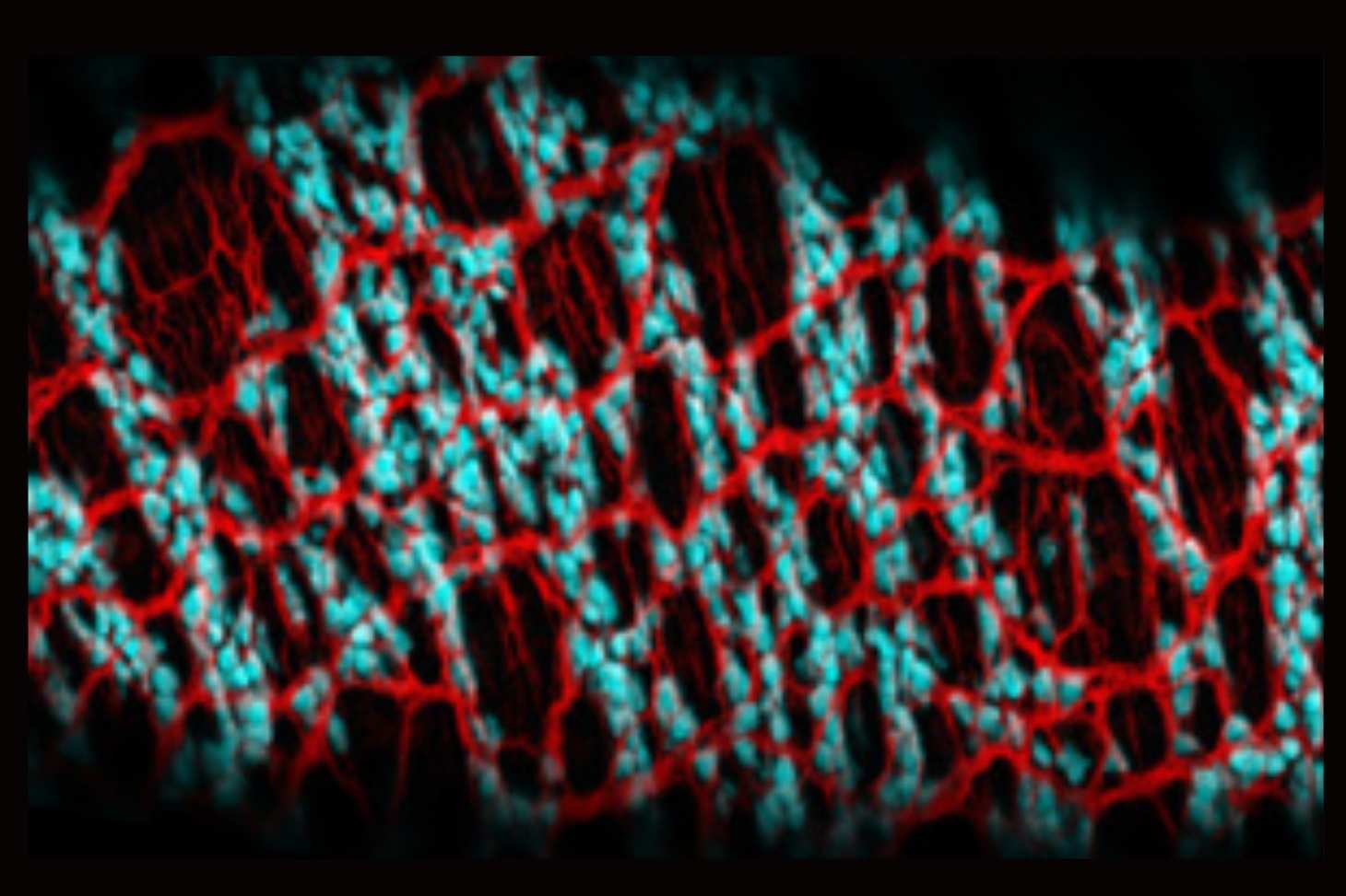
The enteric nervous system, often referred to as the “second brain,” autonomously controls the functions of the digestive tract. A study published in *Genes & Development* reveals that the LKB1 protein plays a key role in the formation of this network by coordinating the differentiation of neurons and glial cells from their embryonic stem cells. This research opens up new avenues for understanding certain congenital diseases and digestive disorders linked to dysfunction of the enteric nervous system.
LKB1, an energy regulator essential for the formation of the "second brain"
The enteric nervous system consists of more than 500 million neurons associated with glial cells that play a role far beyond mere support. They help maintain the intestinal barrier, regulate local immune responses, and control the movements that allow food to move through the digestive tract. These two cell types originate from the same pool of neural crest-derived stem cells during embryonic development.
While the genetic programs that govern this process are now relatively well understood, the influence of cellular metabolism on this stage of development remained largely unexplored. In a study published in the journal
Genes & Development, focused on LKB1, a protein that acts as a sensor for the energy status of cells.
Using a mouse model in which the gene
Lkb1 is specifically deleted in the stem cells that give rise to the enteric nervous system; they demonstrated that the absence of this protein profoundly disrupts the formation of the intestinal nervous network. Neurons fail to differentiate, while glial cells differentiate but gradually degenerate, compromising the integrity of the digestive tissue. These alterations were visualized using high-resolution three-dimensional imaging techniques, combining sheet-light microscopy and adaptive-optics confocal microscopy.
Two distinct mechanisms for neurons and glial cells
Analyses reveal that the loss of LKB1 leads to a significant increase in oxidative stress in the enteric nervous system. This causes DNA damage and activates the p53 protein, a major player in the cellular stress response, leading to the death of many cells.
Scientists have shown, however, that neurons and glial cells do not respond in the same way to this disruption. Inhibiting p53 preserves some of the glial cells but does not restore neuronal differentiation. This observation demonstrates that LKB1 controls the fate of these two cell populations through mechanisms that are at least partially distinct. More than just a cell survival factor, LKB1 thus appears to be a true metabolic checkpoint that directs the neuro-glial fate during development.
Toward a Better Understanding of Diseases of the Enteric Nervous System
The abnormalities observed in mice show similarities to certain human neurocristopathies, notably Hirschsprung’s disease and certain forms of Waardenburg syndrome, which result from abnormal development of neural crest-derived cells. Although no LKB1 mutations have been identified in these patients, this research suggests that a dysfunction in this metabolic pathway could contribute to these conditions or to other enteric neurogliopathies.
Beyond these congenital disorders, the study highlights the central role of enteric glial cells in maintaining digestive health. Their ability to adapt in the face of stress and inflammation makes them promising candidates for future regenerative medicine approaches. A better understanding of the molecular mechanisms that control their development and maintenance could thus open up new therapeutic avenues for diseases of the enteric nervous system.
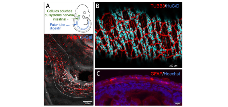
© Florence Appaix, Anthony Lucas, Sakina Torch, Chantal Thibert
Figure: In situ images of neurons and glial cells of the enteric nervous system in the digestive tract. A. Schematic representation of a mouse embryo at 10.5 days of development. The stem cells that colonize the future digestive tract are shown in green. Image of the colonization obtained by Z-stack imaging of the developing digestive tract in the cleared embryo. The neural network is visualized in white using the neuronal marker Peripherin (Periph), and the stem cells are visualized in red using the β-galactosidase (β-Gal) reporter.
B. Image obtained after Z-stack projection of the neural network in the cleared digestive tract of a newborn mouse. The nerve extensions are labeled in red with Tubulin 3 (TUBB3), and the neuron nuclei are labeled in cyan with HuC/D.
C. Cross-section of the intestine showing the glial cells of the enteric nervous system in red (GFAP). All nuclei in the digestive tract are stained blue (Hoechst). These techniques have enabled the detailed characterization of loss-of-function mutants of the LKB1 protein in the stem cells of the enteric nervous system.