

Respiration is central to energy conversion in most living organisms. It relies on electron transport chains located in the membrane, within which small molecules called quinones act as electrochemical shuttles. Among the molecules that fuel these respiratory chains, NADH plays a central role. Produced by numerous metabolic reactions, it must be reoxidized for the cell to continue functioning. This step is carried out by NADH dehydrogenases. Enzymes known as type II, or NDH-II, are widespread throughout the living world. They are generally described as simple flavoproteins capable of binding directly to the membrane to access the quinones. However, this view does not account for their full diversity.
By studying the soil bacterium Bacillus subtilis, scientists, in a paper published in the journal *Nature Communications*, have nevertheless discovered a much more elaborate organization. Using a combination of comparative genomics, genetics, biochemistry, and cryo-electron microscopy, they demonstrated that the Ndh enzyme associates with a partner protein called YjlC, which belongs to the HMP (Helical Membrane Plug-in) protein family.
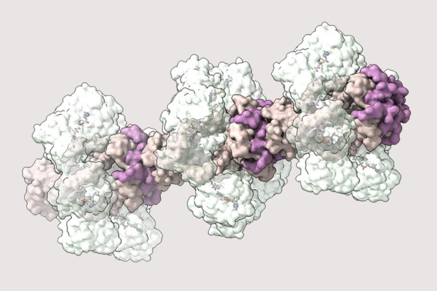
Both proteins are essential for respiratory activity. Four YjlC subunits assemble to form a hydrophobic tube, to which four Ndh enzymes bind. These structures can then stack into fibers that can reach nearly 100 nanometers in length.
Filled with lipids and quinones, this tube functions as an extension of the membrane. It provides the enzyme with organized access to the quinone pool, while reassigning the function of the enzyme’s C-terminal domain: instead of anchoring directly into the bilayer, it serves here as an attachment point for the tube formed by YjlC.
Phylogenetic analyses indicate that this system is unique to the Bacillales. It appears to have resulted from the recruitment, over the course of evolution, of an ancestral HMP module that was already involved in other respiratory complexes.
These results thus call for a reevaluation of the traditional view of NDH-IIs, which have long been considered isolated membrane-associated enzymes. They now appear, at least in certain bacterial lineages, to be components of true supramolecular assemblies specialized in managing electron flow.
Beyond shedding light on the functioning of bacterial energy metabolism, this discovery expands the repertoire of biological architectures used to organize respiratory chains. It also suggests that other, as yet unknown assemblies may exist and offers new avenues for designing organized enzymatic systems for biotechnological applications.
In summary, this study highlights an original strategy for organizing a bacterial NADH dehydrogenase around a lipid tunnel, which lies at the heart of respiration and the energy adaptation of microorganisms.